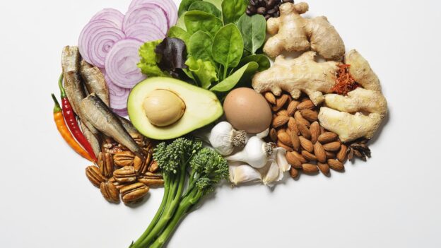
Category Archives: Articles on Brain

Normal Pressure Hydrocephalus

Normal Pressure Hydrocephalus

Normal Pressure Hydrocephalus (NPH) is a neurological disorder that primarily affects older adults. It’s characterized by an abnormal accumulation of cerebrospinal fluid (CSF) in the brain’s ventricles, which can lead to a specific set of symptoms. Despite the name, the CSF pressure is often within the normal range, which can make diagnosis challenging. This comprehensive guide […]

Normal Pressure Hydrocephalus (NPH) is a neurological disorder that primarily affects older adults. It’s characterized by an abnormal accumulation of cerebrospinal fluid (CSF) in the brain’s ventricles, which can lead to a specific set of symptoms. Despite the name, the CSF pressure is often within the normal range, which can make diagnosis challenging. This comprehensive guide aims to provide a detailed understanding of NPH, covering its symptoms, diagnostic processes, and available treatment options.

Understanding Normal Pressure Hydrocephalus

Hydrocephalus, in general, is a condition where there is an excessive accumulation of CSF within the brain. CSF is a clear fluid that surrounds the brain and spinal cord, providing cushioning, nutrient distribution, and waste removal. In NPH, the normal flow and absorption of CSF are disrupted, leading to enlarged ventricles. This enlargement can put pressure on surrounding brain tissue, resulting in a variety of neurological symptoms.

NPH was first described in 1964 by Dr. Salomon Hakim, who identified a unique set of symptoms occurring in patients with normal CSF pressure. It primarily affects individuals over the age of 60, although it can occur in younger adults as well. The condition is often underdiagnosed or misdiagnosed because its symptoms can mimic those of more common disorders like Alzheimer’s disease and Parkinson’s disease.

There are two main types of NPH:

  • Idiopathic NPH: This is the most common form, where the cause of the CSF flow disruption is unknown.
  • Secondary NPH: This type occurs as a result of another underlying condition, such as subarachnoid hemorrhage, meningitis, traumatic brain injury, or previous brain surgery.

The Classic Triad of Symptoms

NPH is characterized by a classic triad of symptoms, often referred to as Hakim’s triad. These include:

  1. Gait Disturbances: This is often the first and most prominent symptom of NPH. Individuals may experience a wide-based, shuffling gait, described as “magnetic gait,” where the feet appear stuck to the floor. They may have difficulty initiating movement, turning, or maintaining balance. The gait disturbance is often slow and unsteady, increasing the risk of falls.
  2. Cognitive Impairment: Cognitive symptoms can range from mild forgetfulness and difficulty concentrating to more significant memory loss and impaired executive function. Patients may struggle with planning, organizing, and problem-solving. The cognitive decline in NPH is often slower and less severe than in Alzheimer’s disease.
  3. Urinary Incontinence: This symptom typically presents as urinary urgency or frequency, progressing to urge incontinence. Patients may have difficulty controlling their bladder, leading to accidental urine leakage. Incontinence can significantly impact a person’s quality of life and social interactions.

It’s important to note that not all individuals with NPH will experience all three symptoms. Some may present with only one or two of the triad, making diagnosis more challenging. The severity of symptoms can also vary significantly from person to person.

Diagnose

Lumbar Puncture (Spinal Tap)

Diagnosing NPH can be complex due to the overlap of symptoms with other neurological conditions. A thorough evaluation by an experienced neurologist is crucial for accurate diagnosis. The diagnostic process typically involves the following steps:

  1. Medical History and Neurological Examination: The neurologist will review the patient’s medical history, including any prior illnesses, injuries, or surgeries. A comprehensive neurological examination will assess gait, balance, coordination, reflexes, cognitive function, and bladder control.
  2. Brain Imaging: Brain imaging techniques, such as MRI and CT scans, play a vital role in diagnosing NPH. These scans can reveal enlarged ventricles and help rule out other conditions that may be causing similar symptoms. MRI is often preferred over CT scans because it provides more detailed images of the brain and can better assess CSF flow.
  3. Cerebrospinal Fluid (CSF) Studies:
    • Lumbar Puncture (Spinal Tap): This procedure involves inserting a needle into the lower spine to collect a sample of CSF. The CSF pressure is measured to confirm that it is within the normal range or only slightly elevated.
    • Large-Volume CSF Tap Test: This test involves removing a larger amount of CSF (30-50 ml) during a lumbar puncture. The patient’s symptoms, particularly gait and cognitive function, are assessed before and after the CSF removal. If symptoms improve significantly after the tap, it suggests that the patient may benefit from shunt surgery.
    • CSF Infusion Test: During this test, fluid is slowly injected into the lower spine while measuring the pressure. If the body cannot absorb the extra fluid, the pressure will rise, which could indicate NPH and that surgery will be helpful.
    • Lumbar Drainage Test: A tube is inserted between the bones in your spine to drain a large amount of CSF over a few days, to see whether your symptoms improve.
  4. Gait Assessment: A licensed physical therapist can perform a gait assessment to quantify the patient’s gait abnormalities and track changes over time. This assessment can help determine the severity of gait impairment and monitor response to treatment.

Treatment Options

Lumboperitoneal (LP) Shunt

The primary treatment for NPH is the surgical placement of a shunt to drain excess CSF from the brain3. There are two main types of shunts:

  1. Ventriculoperitoneal (VP) Shunt: This is the most common type of shunt used to treat NPH. A catheter is inserted into one of the brain’s ventricles and then tunneled under the skin to the abdomen. A valve regulates the flow of CSF from the brain to the abdominal cavity, where it is absorbed.
  2. Lumboperitoneal (LP) Shunt: In this procedure, a catheter is inserted into the lumbar spine and tunneled under the skin to the abdomen. This type of shunt drains CSF from the spinal canal to the abdominal cavity.

Shunt Surgery: The surgery takes about an hour. The surgery typically includes the following5:

  • A small incision followed by a small hole is made in the scalp and skull
  • A tiny hole is made in the meninges (the protective covering of the brain) to allow the placement of a catheter in the lateral ventricle
  • The neurosurgeon will make a few small incisions behind the ear to insert the shunt valve.
  • The catheter is placed under the skin
  • The other end of the catheter will be situated in the abdomen or above the internal jugular vein
  • After the procedure, sterile bandages are placed over each incision

Shunt Complications: While shunt surgery can be highly effective in treating NPH, it’s essential to be aware of potential complications. These can include:

  • Infection: Shunt infections can occur and may require antibiotic treatment or shunt removal.
  • Bleeding: Bleeding in the brain can occur as a result of shunt placement.
  • Shunt Malfunction: Shunts can become blocked or malfunction, requiring revision surgery.
  • Over-drainage or Under-drainage: These can lead to headaches, seizures, or a return of NPH symptoms.

Post-operative care: It’s important that you and your family are aware of these possible complications. If you have this surgery, be sure to follow all of your doctor’s directions and keep any follow-up appointments to make sure it’s working properly.

Alternatives to Shunt Surgery: While shunt surgery is the most common and effective treatment for NPH, other treatment options may be considered in certain cases.

  • Endoscopic Third Ventriculostomy (ETV): This minimally invasive procedure creates a small opening in the floor of the third ventricle, allowing CSF to flow more freely. ETV is typically reserved for patients with obstructive hydrocephalus, where there is a blockage in the CSF pathways.
  • Medical Management: Medications may be used to manage some of the symptoms of NPH, such as bladder control problems or cognitive impairment. However, medication alone cannot address the underlying cause of NPH.

The Importance of Early Diagnosis and Treatment

Early diagnosis and treatment of NPH are crucial for improving patient outcomes. If left untreated, NPH can lead to progressive neurological decline and significant disability. With timely intervention, many individuals with NPH can experience significant improvement in their symptoms and regain a better quality of life.

It is also important to have an accurate diagnosis by an experienced team is essential to getting the best treatment.

Conclusion

Normal Pressure Hydrocephalus is a treatable condition that can significantly impact the lives of older adults. By understanding the symptoms, diagnostic process, and treatment options, individuals and their families can work with healthcare professionals to achieve an early diagnosis and appropriate management.

Shunt surgery remains the most effective treatment for NPH, offering the potential for improved gait, cognitive function, and bladder control. If you or a loved one is experiencing symptoms of NPH, seeking prompt medical attention is essential for optimizing outcomes and enhancing quality of life.

Speak With Us

At Southern California Brain & Spine Surgery, we understand the complexities of neurological conditions like Normal Pressure Hydrocephalus. Our dedicated team of experts is committed to providing comprehensive and personalized care for all your brain and spine health needs. If you or a loved one is experiencing symptoms of NPH or any other neurological concerns, we encourage you to contact us today.

Dr. Moksha's picture

Our experienced neurologists and neurosurgeons will conduct a thorough evaluation, utilize advanced diagnostic techniques, and develop a tailored treatment plan to help you regain your quality of life. Trust Southern California Brain & Spine for compassionate and cutting-edge care. Visit socalbrainspine.com or call us today to schedule a consultation. Your brain and spine health are our top priority.

Intracranial Hypotension

Conservative Management Strategies for Intracranial Hypotension

Intracranial hypotension (IH) is a neurological condition characterized by low cerebrospinal fluid (CSF) pressure, often caused by spinal CSF leaks. Patients typically experience orthostatic headaches, nausea, and cognitive symptoms. While targeted interventions like epidural blood patches remain critical for persistent cases, conservative strategies play a vital role in initial management and symptom relief. This blog […]

Intracranial hypotension (IH) is a neurological condition characterized by low cerebrospinal fluid (CSF) pressure, often caused by spinal CSF leaks. Patients typically experience orthostatic headaches, nausea, and cognitive symptoms. While targeted interventions like epidural blood patches remain critical for persistent cases, conservative strategies play a vital role in initial management and symptom relief. This blog explores evidence-based non-invasive approaches to managing IH, empowering patients and clinicians with practical tools to improve outcomes.

Understanding Conservative Management

Neurosurgeon’s Role in Patient Care, asking second opinion from a neurosurgeon

Conservative strategies aim to alleviate symptoms, reduce CSF loss, and support the body’s natural healing processes. These methods are particularly valuable:

  • As first-line therapy while awaiting diagnostic confirmation
  • For mild cases where symptoms may resolve spontaneously
  • In conjunction with interventional treatments to enhance recovery

Studies suggest 28% of patients with spontaneous IH (SIH) improve with conservative care alone. However, early intervention is crucial to prevent complications like cerebral venous thrombosis or superficial siderosis.

Core Conservative Strategies

1. Bed Rest and Positional Modifications

Bed rest in a supine or Trendelenburg position reduces CSF leakage by lowering hydrostatic pressure on spinal dural defects. Key recommendations:

  • Limit upright activities to <30 minutes initially
  • Sleep with the head slightly elevated (10–30 degrees)
  • Avoid bending, twisting, or lifting >5 lbs

Evidence: A UK consensus guideline emphasizes bed rest as central to symptom relief while awaiting definitive treatment.

2. Hydration and Caffeine

Hydration: Increasing fluid intake (2.5–3 L/day) may boost CSF production. Isotonic beverages like electrolyte solutions are preferred over plain water.
Caffeine: Acts as a cerebral vasoconstrictor and adenosine receptor antagonist, potentially reducing headache severity.

  • Oral: 100–300 mg every 4–6 hours (max 1.2 g/day)
  • IV caffeine (500 mg) for severe acute symptoms

Limitations: While caffeine provides temporary relief, prolonged use can lead to tolerance and rebound headaches.

3. Abdominal Binders

Compression garments (20–30 mmHg pressure) increase intra-abdominal pressure, counteracting CSF loss. Patients report:

  • Delayed headache onset when upright
  • Improved tolerance for brief standing/walking

Protocol: Wear during waking hours, adjusting tightness for comfort.

4. Pharmacological Support

MedicationRoleConsiderations
NSAIDsReduce inflammationAvoid in renal impairment
AcetaminophenMild analgesiaMax 3 g/day
GabapentinNeuropathic painStart low (100 mg TID)
TheophyllineAdenosine blockadeMonitor serum levels

Note: Opioids are discouraged due to dependency risks.

5. Nutritional Optimization

Emerging evidence highlights dietary strategies to support CSF production and tissue repair 3:

  • High-sodium foods: Olives, broth, pickles (aim for 3–5 g/day)
  • Omega-3 fatty acids: Fatty fish, walnuts, flaxseeds (anti-inflammatory)
  • Magnesium-rich foods: Spinach, almonds, dark chocolate (nerve function)
  • Vitamin B12: Eggs, fortified cereals (myelin sheath repair)

Avoid: Alcohol, caffeine excess, and processed sugars that exacerbate dehydration.

Integrative Approaches

Physical Activity Modifications

While strenuous exercise is prohibited, structured movement prevents deconditioning:

  • Pool walking: Hydrostatic pressure counteracts CSF hypotension.
  • Recumbent cycling: Maintains cardiovascular fitness without spinal strain.
  • Balance training: Seated yoga or tai chi enhances proprioception.

Avoid: Activities involving spinal rotation, bouncing, or Valsalva maneuvers.

Mind-Body Techniques

Chronic pain and fatigue require holistic management:

  • Guided meditation: Reduces stress-induced cortisol spikes.
  • Paced breathing: Improves autonomic dysregulation common in IH.
  • Cognitive behavioral therapy (CBT): Addresses anxiety/helplessness.

Supplements

SupplementProposed BenefitDosage
Coenzyme Q10Mitochondrial energy production100–300 mg/day
Vitamin D3Bone/cartilage health2,000–5,000 IU/day
Turmeric (Curcumin)Anti-inflammatory500 mg BID

Caution: Consult providers to avoid interactions with prescribed therapies.

When to Transition to Interventional Treatments

Conservative measures should not delay definitive care if:

  • Symptoms persist >2 weeks
  • Neurological deficits emerge (e.g., diplopia, tinnitus)
  • MRI shows progressive complications (subdural hematoma)

First-line intervention: Non-targeted epidural blood patches (20–30 mL autologous blood) resolve 72% of cases within two procedures.

Conclusion

Conservative management for intracranial hypotension combines physiological support, symptom modulation, and lifestyle adaptations. While these strategies provide crucial relief, they work best within a multidisciplinary framework. Early collaboration with neurologists and interventional radiologists ensures timely escalation to advanced therapies when needed.

Why Choose Southern California Brain & Spine Surgery?

While this blog focuses on intracranial hypotension, traumatic skull fractures and brain injuries demand specialized neurosurgical expertise. At Southern California Brain & Spine Surgery, our team led by Dr. Moksha Ranasinghe offers:

  • Advanced diagnostics: High-resolution CT/MRI for precise fracture assessment
  • Minimally invasive repairs: Endoscopic techniques for complex cranial defects
  • Post-traumatic rehabilitation: Customized plans addressing CSF leaks or secondary IH
Neurosurgeon Los Angeles - Dr. Moksha Ranasinghe

If you or a loved one has sustained head trauma, early intervention prevents life-threatening complications. Visit socalbrainspine.com or call (213) 369-4583 to schedule a consultation with Los Angeles’ leading neurosurgical team.

Intracranial Hypotension

Understanding Intracranial Hypotension: Causes, Symptoms, and Treatments

Intracranial hypotension (IH) is a medical condition characterized by abnormally low pressure within the skull due to a reduction in cerebrospinal fluid (CSF). This fluid is crucial for protecting the brain and spinal cord, providing cushioning and facilitating nutrient transport. When there is a decrease in CSF volume, it can lead to various neurological symptoms […]

Intracranial hypotension (IH) is a medical condition characterized by abnormally low pressure within the skull due to a reduction in cerebrospinal fluid (CSF). This fluid is crucial for protecting the brain and spinal cord, providing cushioning and facilitating nutrient transport. When there is a decrease in CSF volume, it can lead to various neurological symptoms and complications. This article aims to provide a comprehensive overview of intracranial hypotension, including its causes, symptoms, diagnosis, and treatment options.

What is Intracranial Hypotension?

Intracranial Hypotension

Intracranial hypotension occurs when the pressure of cerebrospinal fluid drops below normal levels, typically below 50 mmHg. This condition can arise from several factors, primarily involving CSF leaks. The most common form of intracranial hypotension is spontaneous intracranial hypotension (SIH), which occurs without an obvious cause, often due to weak spots in the dura mater—the protective layer surrounding the brain and spinal cord.

Causes

Intracranial hypotension can be classified into two main categories: spontaneous and secondary.

  1. Spontaneous Intracranial Hypotension (SIH):
    • SIH typically results from small tears in the dura mater that allow CSF to leak out. These tears can occur due to trivial trauma or may be associated with connective tissue disorders such as Ehlers-Danlos syndrome or Marfan syndrome.
    • In some cases, patients may not recall any specific injury that led to the leak.
  2. Secondary Intracranial Hypotension:
    • This type is often a result of medical procedures or conditions that compromise the integrity of the dura mater. Common causes include:
      • Lumbar punctures: A spinal tap can inadvertently cause a CSF leak.
      • Epidural anesthesia: The placement of an epidural can lead to dural puncture.
      • Spinal surgery or trauma: Procedures involving the spine may result in damage to the dura.
      • Over-drainage from shunt systems: Patients with hydrocephalus may experience IH if their shunt drains too much CSF.

Symptoms

Sensitivity to light or sound

The hallmark symptom is a positional headache, which typically worsens when sitting or standing and improves when lying down. Other symptoms may include:

  • Sensitivity to light or sound
  • Nausea, with or without vomiting
  • Neck pain or stiffness
  • Hearing changes, such as ringing in the ears
  • Cognitive difficulties, including trouble concentrating
  • Double vision
  • Dizziness and imbalance
  • Memory impairment

These symptoms can significantly impact daily life and may lead individuals to seek medical attention.

Diagnosis

Diagnosing intracranial hypotension involves a combination of clinical evaluation and imaging studies:

  1. Clinical Examination:
    • A thorough physical and neurological examination is essential for assessing symptoms and ruling out other conditions.
  2. Imaging Techniques:
    • Magnetic Resonance Imaging (MRI): MRI is critical for diagnosing IH. It can reveal characteristic findings such as pachymeningeal enhancement (thickening of the meninges), sagging of the brain, and subdural fluid collections.
    • Myelography: This technique involves injecting contrast dye into the spinal canal to identify leaks.
  3. Cerebrospinal Fluid Analysis:
    • A lumbar puncture may be performed to measure opening pressure and analyze CSF composition. In IH cases, CSF pressure is typically low, with elevated protein levels.

Treatment Options

Epidural Blood Patch

Treatment focuses on alleviating symptoms and addressing the underlying cause:

  1. Conservative Management:
    • Many patients experience symptom relief through conservative measures such as:
      • Bed rest: Reducing activity can help alleviate headaches.
      • Increased fluid intake: Staying hydrated supports CSF production.
      • Caffeine consumption: Caffeine can temporarily raise CSF pressure.
      • Pain relievers: Over-the-counter medications may help manage headache pain.
  2. Epidural Blood Patch:
    • If conservative treatments are ineffective, an epidural blood patch may be recommended. This procedure involves injecting a small amount of the patient’s blood into the epidural space to seal the leak and restore normal CSF pressure.
  3. Surgical Intervention:
    • In cases where a dural tear is identified, surgical repair may be necessary to address the source of the leak directly.
  4. Management of Underlying Conditions:
    • If IH results from over-drainage of a shunt system, adjusting or replacing the shunt valve may be required.

Conclusion

Intracranial hypotension is a complex condition that can significantly affect quality of life due to its debilitating symptoms. Understanding its causes, recognizing its symptoms, and seeking appropriate treatment are crucial for effective management. If you or someone you know is experiencing symptoms associated with intracranial hypotension, it’s essential to consult a healthcare professional for an accurate diagnosis and tailored treatment plan.

Neurosurgeon Los Angeles, Spine Surgeon in Los Angeles.

For expert care regarding brain, skull, and spine fractures or any concerns related to intracranial conditions, visit Southern California Brain & Spine Surgery. Our dedicated team specializes in providing comprehensive evaluations and advanced treatment options tailored to your needs. Don’t wait—take control of your health today!

Spinal AVMs

Preventing Complications from Arteriovenous Malformations (AVM)

Arteriovenous malformations (AVMs) are abnormal connections between arteries and veins that disrupt normal blood flow. These complex vascular anomalies can occur in various parts of the body, including the brain and spinal cord, and may lead to serious complications such as hemorrhage, neurological deficits, and chronic pain. Understanding how to prevent complications associated with AVMs […]

Arteriovenous malformations (AVMs) are abnormal connections between arteries and veins that disrupt normal blood flow. These complex vascular anomalies can occur in various parts of the body, including the brain and spinal cord, and may lead to serious complications such as hemorrhage, neurological deficits, and chronic pain. Understanding how to prevent complications associated with AVMs is crucial for those diagnosed with this condition. This blog will explore the nature of AVMs, their potential complications, and effective strategies for prevention and management.

Understanding Arteriovenous Malformations

What Are AVMs?

Causes of Arteriovenous Malformations

AVMs are congenital malformations characterized by a tangle of blood vessels where arterial blood flows directly into the venous system without passing through capillaries. This abnormal connection can lead to increased blood flow and pressure in the veins, which may cause them to enlarge or rupture over time. While some individuals with AVMs remain asymptomatic throughout their lives, others may experience significant health issues.

Types of AVMs

AVMs can be classified based on their location:

  1. Cerebral AVMs: Located in the brain, these are particularly concerning due to the risk of bleeding.
  2. Spinal AVMs: Found in the spinal cord, they can lead to neurological symptoms.
  3. Peripheral AVMs: Occurring in the limbs or other body areas, these can cause pain and swelling.

Potential Complications of AVMs

Heart failure cause of AVM

The most severe complication associated with AVMs is hemorrhage. The risk of bleeding varies depending on factors such as the size and location of the AVM. Studies indicate that unruptured brain AVMs have an annual bleeding risk of approximately 2-4%. Other complications may include:

  • Neurological deficits: Damage to surrounding brain tissue can result in seizures, headaches, or cognitive impairments.
  • Pain and swelling: Peripheral AVMs may cause chronic pain and swelling in affected limbs.
  • Heart failure: High-output cardiac failure can occur due to increased blood flow demands on the heart from large AVMs.

Strategies for Preventing Complications

While it is not possible to prevent the formation of AVMs, several strategies can help manage existing conditions and reduce the risk of complications.

1. Regular Monitoring

For individuals diagnosed with an AVM, regular monitoring through imaging studies (such as MRI or angiography) is essential. This allows healthcare providers to track changes in size or symptoms over time. If an AVM shows signs of growth or increased risk of bleeding, timely intervention may be necessary.

2. Prompt Treatment

If symptoms develop or worsen, seeking prompt medical attention is critical. Treatments for AVMs include:

  • Embolization: A minimally invasive procedure where materials are injected into the blood vessels supplying the AVM to reduce blood flow. This can help shrink the malformation and lower bleeding risks.
  • Surgery: In some cases, surgical resection may be recommended to remove the AVM entirely, especially if it poses a high risk of bleeding.
  • Radiation therapy: Stereotactic radiosurgery uses focused radiation beams to damage the abnormal blood vessels over time, leading to closure.

3. Lifestyle Modifications

Certain lifestyle changes can help manage symptoms and reduce risks associated with AVMs:

  • Avoiding high-impact activities: Individuals with cerebral AVMs should avoid contact sports or activities that could lead to head injury.
  • Managing stress: High-stress levels can exacerbate symptoms; practices such as meditation or yoga may help.
  • Healthy diet and exercise: Maintaining a balanced diet and engaging in low-impact exercise can promote overall vascular health.

4. Medication Management

Medications may be prescribed to manage symptoms related to AVMs, such as headaches or seizures. Regular consultations with healthcare providers ensure that medications are effective and adjusted as needed.

5. Education and Support

Understanding your condition is vital for effective management. Patients should educate themselves about their specific type of AVM, potential symptoms of complications, and treatment options available. Support groups can also provide valuable resources and emotional support for individuals living with AVMs.

Conclusion

Arteriovenous malformations present unique challenges that require careful management to prevent serious complications. By engaging in regular monitoring, seeking prompt treatment when necessary, making lifestyle modifications, managing medications effectively, and educating oneself about the condition, individuals with AVMs can significantly reduce their risk of adverse outcomes.

Neurosurgeon Los Angeles, Spine Surgeon in Los Angeles.

For those seeking expert guidance on managing arteriovenous malformations or exploring treatment options tailored to their specific needs, visiting Southern California Brain & Spine is highly recommended. Their team specializes in vascular conditions like AVMs and offers a comprehensive approach that combines advanced technology with compassionate care. Ensuring you have access to knowledgeable professionals can make a significant difference in your treatment journey and overall quality of life.

Chiari Malformation

Chiari Malformation: Symptoms, Diagnosis, and Treatment Options

Chiari malformation is a neurological condition characterized by the displacement of brain tissue into the spinal canal. This condition can lead to a variety of symptoms, ranging from mild discomfort to severe neurological issues. Understanding the symptoms, diagnosis, and treatment options available is crucial for those affected by this condition. What is Chiari Malformation? Chiari […]

Chiari malformation is a neurological condition characterized by the displacement of brain tissue into the spinal canal. This condition can lead to a variety of symptoms, ranging from mild discomfort to severe neurological issues. Understanding the symptoms, diagnosis, and treatment options available is crucial for those affected by this condition.

What is Chiari Malformation?

Chiari malformation occurs when the cerebellum, the part of the brain that controls balance, extends into the spinal canal. This can disrupt the normal flow of cerebrospinal fluid (CSF), leading to increased pressure on the brain and spinal cord. There are several types of Chiari malformation, with Type I being the most common. While some individuals may remain asymptomatic, others may experience significant health challenges.

Symptoms of Chiari Malformation

The symptoms of Chiari malformation can vary widely depending on the type and severity of the condition. Common symptoms include:

  • Headaches: Often triggered by coughing, sneezing, or straining, headaches are one of the most prevalent symptoms.
  • Neck Pain: Discomfort or pain in the neck area can occur, often radiating to the shoulders.
  • Dizziness and Balance Issues: Patients may experience episodes of dizziness, unsteadiness, and coordination problems.
  • Numbness or Tingling: Some individuals report sensations of numbness or tingling in the arms and legs.
  • Vision Problems: Blurred or double vision can also be a symptom.
  • Difficulty Swallowing: This can occur due to pressure on the brainstem.
  • Weakness: Muscle weakness, particularly in the arms, may be experienced.

Symptoms can manifest at any age, but they often become noticeable during late childhood or adulthood. It is important to consult a healthcare professional if any of these symptoms develop, as they may resemble other medical conditions.

Diagnosis of Chiari Malformation

Diagnosing Chiari malformation typically involves a thorough medical history, physical examination, and imaging tests. The following diagnostic methods are commonly used:

  • Magnetic Resonance Imaging (MRI): This is the most effective imaging technique for diagnosing Chiari malformation. An MRI provides detailed images of the brain and spinal cord, allowing doctors to assess the extent of the malformation.
  • Computerized Tomography (CT) Scan: A CT scan can help visualize structural abnormalities in the brain and skull.
  • X-rays: These may be used to rule out other potential causes of symptoms.
MRI of Chiari Malformation

During the diagnostic process, healthcare providers will evaluate symptoms, medical history, and any potential family history of neurological conditions. The combination of clinical evaluation and imaging studies helps in confirming the diagnosis of Chiari malformation.

Treatment Options for Chiari Malformation

Treatment for Chiari malformation depends on the severity of symptoms and the impact on the patient’s quality of life. Options may include:

1. Observation and Monitoring

Doctors may recommend a “watchful waiting” approach for individuals with mild symptoms or those who are asymptomatic. Regular follow-up appointments and MRI scans can help monitor the condition over time.

2. Medications

When symptoms like headaches or pain occur, doctors may prescribe medications to manage discomfort. Pain relievers, muscle relaxants, or anti-inflammatory drugs can provide symptomatic relief.

3. Surgery

When symptoms are severe or progressive, surgical intervention may be necessary. The most common surgical procedure for Chiari malformation is posterior fossa decompression. This surgery involves removing a small section of bone from the back of the skull to relieve pressure on the brain and spinal cord. The dura mater, the protective covering of the brain, may also be opened and patched to create additional space for CSF flow.

Other surgical options may include:

  • Spinal Laminectomy: This procedure involves removing part of the bony arch of the spinal canal to alleviate pressure on the spinal cord.
  • Electrocautery: This technique uses high-frequency electrical currents to shrink the lower part of the cerebellum.

A neurosurgeon plays a critical role in the treatment of Chiari malformation. Specialized medical doctors perform surgeries to relieve pressure on the brain and spinal cord, restore normal cerebrospinal fluid flow, and address any associated complications. Consulting a neurosurgeon is essential for determining the most appropriate treatment plan based on the individual’s specific condition and symptoms.

Conclusion

Chiari malformation is a complex neurological condition that can significantly impact a person’s quality of life. Understanding the symptoms, diagnosis, and treatment options is vital for those affected. If you or a loved one is experiencing symptoms associated with Chiari malformation, it is crucial to seek medical advice from a qualified healthcare professional.

Contact Us

Neurosurgeon Los Angeles, Spine Surgeon in Los Angeles.

For expert evaluation and personalized treatment options, we encourage you to contact Southern California Brain & Spine. Our team of experienced neurosurgeons is dedicated to providing comprehensive care tailored to your needs. Don’t let Chiari malformation control your life—reach out to us today and take the first step towards relief and recovery. Visit socalbrainspine.com to learn more about our services and schedule a consultation.

Cerebrovascular Disease

Recognizing the Signs: Early Detection of Cerebrovascular Events

Cerebrovascular disease encompasses a range of conditions that affect blood flow to the brain, leading to serious health issues such as strokes, transient ischemic attacks (TIAs), and brain aneurysms. Early detection of these events is crucial for effective treatment and can significantly improve outcomes for patients. This blog aims to educate readers on the signs […]

Cerebrovascular disease encompasses a range of conditions that affect blood flow to the brain, leading to serious health issues such as strokes, transient ischemic attacks (TIAs), and brain aneurysms. Early detection of these events is crucial for effective treatment and can significantly improve outcomes for patients. This blog aims to educate readers on the signs of cerebrovascular events, the importance of prompt medical attention, and preventive measures that can be taken.

Understanding Cerebrovascular Disease

Cerebrovascular disease refers to any condition that affects the blood vessels in the brain. The most common types include:

Cerebrovascular Disease
  • Stroke: This occurs when blood flow to a part of the brain is interrupted, either due to a blockage (ischemic stroke) or a rupture (hemorrhagic stroke).
  • Transient Ischemic Attack (TIA): Often referred to as a “mini-stroke,” a TIA is a temporary period of symptoms similar to those of a stroke. TIAs are often warning signs that a full-blown stroke may occur in the future.
  • Brain Aneurysm: This is a bulge in a blood vessel in the brain that can leak or rupture, leading to bleeding in the brain.
  • Carotid Artery Disease: Narrowing of the carotid arteries can reduce blood flow to the brain and increase the risk of stroke.

According to the Cleveland Clinic, cerebrovascular disease is a leading cause of death in the United States, responsible for over 160,000 deaths annually. Understanding its signs and symptoms can save lives.

Recognizing Early Signs of Cerebrovascular Events

The American Stroke Association emphasizes using the acronym F.A.S.T. to help identify potential strokes quickly:

  • Face drooping: Ask the person to smile. Does one side of their face droop or feel numb?
  • Arm weakness: Ask them to raise both arms. Does one arm drift downward?
  • Speech difficulty: Ask them to repeat a simple phrase. Is their speech slurred or strange?
  • Time to call emergency services: If you observe any of these signs, call 911 immediately.

In addition to these classic signs, other symptoms may include:

  • Sudden confusion or trouble understanding speech
  • Sudden trouble seeing in one or both eyes
  • Sudden trouble walking, dizziness, loss of balance, or coordination
  • Severe headache with no known cause

Recognizing these symptoms early can make a significant difference in treatment outcomes. The faster medical intervention occurs, the better the chances for recovery and minimizing long-term damage.

The Importance of Timely Medical Intervention

Cerebrovascular events can lead to irreversible brain damage within minutes if not treated promptly. For instance, during an ischemic stroke, brain cells begin dying within three hours due to lack of oxygen and nutrients4. Therefore, immediate medical attention is crucial.

Medical professionals typically perform several diagnostic tests upon arrival at an emergency room:

Neurosurgeon’s Role in Patient Care, asking second opinion from a neurosurgeon

  1. CT Scan: This imaging test helps determine whether a stroke is ischemic or hemorrhagic.
  2. MRI: Magnetic resonance imaging provides detailed images of brain tissue and can identify areas affected by a stroke.
  3. Blood Tests: These tests assess blood clotting factors and other health markers.
  4. Carotid Ultrasound: This test evaluates blood flow through the carotid arteries.

Treatment options vary depending on whether the stroke is ischemic or hemorrhagic:

  • For ischemic strokes, treatments may include clot-busting medications like tPA (tissue plasminogen activator) if administered within a specific time frame.
  • Hemorrhagic strokes may require surgical intervention to repair blood vessels or relieve pressure on the brain.

Risk Factors for Cerebrovascular Disease

Understanding risk factors is essential for prevention:

Non-Modifiable Risk Factors

  • Age: Risk increases with age.
  • Family history: A family history of cerebrovascular diseases increases risk.
  • Race and gender: Certain racial groups and males are at higher risk.

Modifiable Risk Factors

  • Hypertension: High blood pressure is one of the most significant risk factors for stroke.
  • Diabetes: Poorly controlled diabetes increases stroke risk.
  • Smoking: Tobacco use significantly raises the likelihood of cerebrovascular events.
  • High cholesterol levels: Elevated cholesterol can lead to artery blockage.
  • Obesity and sedentary lifestyle: Lack of physical activity contributes to many health issues.

By addressing modifiable risk factors through lifestyle changes—such as maintaining a healthy diet, exercising regularly, managing stress, and avoiding tobacco use—individuals can significantly reduce their risk of cerebrovascular disease.

Preventive Measures

Preventing cerebrovascular disease involves proactive health management:

  1. Regular Check-ups: Routine visits with healthcare providers can help monitor blood pressure, cholesterol levels, and overall health.
  2. Healthy Diet: A diet rich in fruits, vegetables, whole grains, lean proteins, and healthy fats can improve cardiovascular health.
  3. Physical Activity: Engaging in regular physical activity helps maintain healthy body weight and reduces hypertension and diabetes risk.
  4. Stress Management: Techniques such as mindfulness meditation, yoga, or deep-breathing exercises can help manage stress levels effectively.
  5. Medication Adherence: For those with existing conditions like hypertension or diabetes, adhering to prescribed medications is crucial.

Conclusion

Recognizing the early signs of cerebrovascular events is vital for effective treatment and recovery. By understanding symptoms such as facial drooping, arm weakness, speech difficulties, and other warning signs outlined by F.A.S.T., individuals can act swiftly in emergencies.

Neurosurgeon Los Angeles - Dr. Moksha Ranasinghe

Cerebrovascular disease poses significant risks but is largely preventable through lifestyle modifications and regular health monitoring. If you or someone you know exhibits symptoms suggestive of a cerebrovascular event, seek immediate medical attention.

For expert care in managing cerebrovascular conditions or if you have concerns about your risk factors, contact Southern California Brain & Spine Surgery today. Our dedicated team specializes in diagnosing and treating cerebrovascular diseases with advanced techniques tailored to your needs. Don’t wait—your health matters!

Chiari Malfunction

How is Chiari Malformation Diagnosed?

Chiari Malformation is a neurological condition where brain tissue extends into the spinal canal, often leading to various symptoms. Understanding how this condition is diagnosed is crucial for those experiencing related symptoms or for those who have been diagnosed with it. This blog will delve into the diagnostic process for Chiari Malformation, including the types, […]

Chiari Malformation is a neurological condition where brain tissue extends into the spinal canal, often leading to various symptoms. Understanding how this condition is diagnosed is crucial for those experiencing related symptoms or for those who have been diagnosed with it. This blog will delve into the diagnostic process for Chiari Malformation, including the types, symptoms, and the importance of accurate diagnosis.

What is Chiari Malformation?

Chiari Malfunction

Chiari Malformation is classified into several types, with Type I being the most common. In Type I, the lower part of the cerebellum (the brain region responsible for coordination and balance) extends through the foramen magnum, the opening at the base of the skull. Other types include Chiari II (associated with spina bifida), Chiari III (involves herniation of brain tissue), and Chiari IV (a rare condition where part of the cerebellum does not develop properly).

This condition can be congenital (present at birth) or acquired later in life due to factors such as injury or tumors. While some individuals may remain asymptomatic throughout their lives, others may experience debilitating symptoms that require medical intervention.

Symptoms of Chiari Malformation

Symptoms can vary widely among individuals and may include:

  • Headaches: Often occurring at the back of the head, these can worsen with coughing or sneezing.
  • Neck pain: Discomfort or pain in the neck area.
  • Balance issues: Difficulty maintaining balance or coordination.
  • Numbness: Tingling or loss of sensation in arms and legs.
  • Vision problems: Such as blurred vision or double vision.
  • Difficulty swallowing: Known as dysphagia.
  • Fatigue: General tiredness that can affect daily activities.

These symptoms can resemble other conditions, making diagnosis challenging. Therefore, a thorough evaluation by a healthcare professional is essential.

The Diagnostic Process

The diagnosis of Chiari Malformation typically involves several steps:

  1. Medical History Review: The physician will begin by taking a comprehensive medical history. This includes discussing any symptoms experienced, their onset, and any family history of neurological disorders.
  2. Physical Examination: A neurological examination will be conducted to assess various functions such as balance, coordination, reflexes, and sensory perception. This helps identify any neurological deficits that may suggest Chiari Malformation.
  3. Imaging Tests:
    • Magnetic Resonance Imaging (MRI): This is the primary diagnostic tool used to confirm Chiari Malformation. An MRI provides detailed images of the brain and spinal cord, allowing doctors to visualize abnormalities such as cerebellar tonsil herniation. In cases where patients cannot undergo an MRI, alternative tests like myelography may be employed.
    • Cine MRI: A specialized form of MRI that evaluates cerebrospinal fluid (CSF) flow dynamics around the brain and spinal cord can help determine if there is obstruction caused by the malformation.
    • CT Scans and X-rays: These imaging techniques may also be utilized to assess bony abnormalities at the base of the skull and cervical spine that could contribute to symptoms.
  4. Additional Tests: In some cases, further assessments may be necessary to evaluate associated conditions like syringomyelia (the formation of a cyst within the spinal cord), which can complicate Chiari Malformation.

Challenges in Diagnosis

Symptoms of Arteriovenous Malformations

Diagnosing Chiari Malformation can be complex due to overlapping symptoms with other conditions such as migraines, multiple sclerosis, or even anxiety disorders. Many patients are diagnosed incidentally during imaging studies performed for unrelated issues. Therefore, if you suspect you have Chiari Malformation or are experiencing relevant symptoms, seeking a specialist’s opinion is crucial.

Importance of Early Diagnosis

Early diagnosis is vital for effective management and treatment of Chiari Malformation. If left untreated, symptoms can worsen over time and lead to significant complications such as chronic pain, mobility issues, and neurological deficits. For symptomatic individuals, timely intervention can greatly improve quality of life.

Conclusion

Chiari Malformation is a complex condition requiring careful evaluation and diagnosis. The diagnostic process involves a detailed medical history review, physical examination, and advanced imaging techniques like MRI to confirm the presence of abnormalities. If you experience symptoms associated with Chiari Malformation or have concerns about your neurological health, it is essential to consult a qualified healthcare provider.

Neurosurgeon Los Angeles - Dr. Moksha Ranasinghe

At SoCal Brain & Spine Institute, we specialize in diagnosing and treating conditions like Chiari Malformation. Our team of experienced professionals uses state-of-the-art technology and personalized care plans to ensure you receive the best possible treatment tailored to your needs. Don’t wait for your symptoms to worsen—schedule an appointment with us today to take control of your health! Your well-being is our priority.

Ganglioglioma vs. Other Brain Tumors: Key Differences Explained

Gangliogliomas are a unique type of brain tumor that blend neuronal and glial cells, making them distinct from other brain tumors. Understanding their characteristics, symptoms, treatment options, and how they differ from other brain tumors can be crucial for patients and their families. This blog will explore gangliogliomas in depth, comparing them with other common […]

Gangliogliomas are a unique type of brain tumor that blend neuronal and glial cells, making them distinct from other brain tumors. Understanding their characteristics, symptoms, treatment options, and how they differ from other brain tumors can be crucial for patients and their families. This blog will explore gangliogliomas in depth, comparing them with other common brain tumors to provide clarity on this complex topic.

What is Ganglioglioma?

Ganglioglioma

Ganglioglioma is classified as a low-grade glioma, characterized by the presence of both dysplastic ganglion cells and neoplastic glial cells. These tumors are most commonly located in the temporal lobe of the brain, particularly affecting children and young adults, where they are frequently associated with epileptic seizures. Although gangliogliomas account for only about 2% of all primary brain tumors, they can arise in various locations throughout the central nervous system, including the spinal cord.

The slow-growing nature of gangliogliomas often leads to a better prognosis compared to high-grade gliomas, as they tend to be less aggressive and are more amenable to surgical intervention. In many cases, complete surgical resection can lead to a cure, particularly when the tumor is accessible and can be removed entirely. However, if the tumor cannot be fully excised due to its location or other factors, treatment may involve chemotherapy or radiation therapy.

The symptoms of ganglioglioma can vary based on tumor size and location but often include headaches, nausea, and neurological deficits. Overall, while gangliogliomas are rare, they represent an important category of brain tumors that require careful management and monitoring due to their potential impact on quality of life and neurological function.

Types of Brain Tumors

To understand ganglioglioma better, it is essential to compare it with other types of brain tumors. Here are some common types:

Astrocytomas

Astrocytomas
  • Composed primarily of astrocytes (glial cells).
  • Can be low-grade (like pilocytic astrocytoma) or high-grade (like glioblastoma).
  • Symptoms vary based on location but may include headaches and seizures.

Oligodendrogliomas

Oligodendrogliomas
  • Arise from oligodendrocytes and are generally slow-growing.
  • Often found in the frontal and temporal lobes.
  • Patients may experience seizures or neurological deficits.

Ependymomas

Ependymomas
  • Originates from ependymal cells lining the ventricles of the brain or spinal cord.
  • Symptoms may include headaches, nausea, and changes in vision depending on location.

Meningiomas

Meningiomas
  • Develop from the meninges (the protective layers around the brain).
  • Typically slow-growing and often benign.
  • Symptoms depend on size and location but may include headaches or seizures.

Medulloblastomas

  • Primarily found in children, arising from cerebellar cells.
  • Often aggressive and classified as a high-grade tumor.
  • Symptoms may include headaches, balance issues, and vomiting.

Key Differences Between Ganglioglioma and Other Brain Tumors

Histological Characteristics

Gangliogliomas are unique due to their mixed cellular composition. They contain both glial cells and ganglion cells, which is not seen in most other types of brain tumors that typically consist solely of either glial or neuronal components. This distinct histology influences their behavior, prognosis, and treatment approaches.

Growth Rate

Gangliogliomas are generally slow-growing tumors. Most cases are classified as WHO grade I, indicating a lower likelihood of recurrence after complete surgical resection. In contrast, many other brain tumors can be aggressive (e.g., glioblastoma) or have a higher potential for recurrence even after treatment.

Genetic Features

A significant portion of gangliogliomas harbor specific genetic mutations, particularly in the BRAF gene (specifically the p.V600E mutation), which can influence treatment options. While genetic mutations also play a role in other tumor types (e.g., IDH mutations in astrocytomas), gangliogliomas have a more defined genetic profile that can guide targeted therapies.

Treatment Approaches

Treatment for gangliogliomas often involves surgical resection as the first line of defense. In many cases, complete removal can lead to long-term remission3. Other brain tumors may require a combination of surgery, chemotherapy, and radiation therapy depending on their grade and location:

  • Astrocytomas may require aggressive treatment including chemotherapy and radiation if they are high-grade.
  • Meningiomas, while often benign, may still necessitate surgical intervention if symptomatic or large.
  • Medulloblastomas typically require multimodal treatment due to their aggressive nature.

Diagnosis of Ganglioglioma

Diagnosing ganglioglioma involves several steps:

  1. Imaging Studies: MRI is crucial for identifying the tumor’s location and characteristics. Gangliogliomas often appear as cystic lesions with solid components on imaging studies2.
  2. Biopsy: A definitive diagnosis usually requires a biopsy to analyze tissue samples histologically.
  3. Genetic Testing: Testing for specific mutations like BRAF can provide insights into prognosis and treatment options.

Prognosis

The prognosis for patients with ganglioglioma is generally favorable compared to many other brain tumors. When completely resected, patients often experience long-term survival without recurrence. However, those with anaplastic gangliogliomas (high-grade variants) may face more significant challenges regarding treatment outcomes and overall survival.

Conclusion

Understanding ganglioglioma in comparison to other brain tumors highlights its unique characteristics—both clinically and histologically. While it shares some symptoms with other tumors, its mixed cellular composition, genetic features, and typically favorable prognosis set it apart from more aggressive forms like glioblastoma or medulloblastoma.

Neurosurgeon Los Angeles - Dr. Moksha Ranasinghe

If you or a loved one is facing a diagnosis related to ganglioglioma or any brain tumor condition, it’s essential to seek specialized care. At Southern California Brain & Spine Surgery, our team of experts is dedicated to providing comprehensive evaluations and personalized treatment plans tailored to your specific needs. Don’t hesitate—schedule an appointment today to ensure you receive the best possible care for your condition!

Gangliogliomas

The Importance of Early Detection in Ganglioglioma Treatment

Gangliogliomas are rare brain tumors that predominantly affect children and young adults. Though classified as low-grade tumors, they can lead to significant neurological issues and may transform into higher-grade, more aggressive forms if not treated promptly. This blog explores the critical role of early detection in the treatment of gangliogliomas, outlining the benefits of timely […]

Gangliogliomas are rare brain tumors that predominantly affect children and young adults. Though classified as low-grade tumors, they can lead to significant neurological issues and may transform into higher-grade, more aggressive forms if not treated promptly. This blog explores the critical role of early detection in the treatment of gangliogliomas, outlining the benefits of timely intervention, the challenges faced in diagnosis, and the implications for patient outcomes.

Understanding Gangliogliomas

Gangliogliomas

Gangliogliomas are a type of neuroepithelial tumor characterized by a combination of neuronal and glial cells. They typically occur in the temporal lobes of the brain but can appear in various locations. These tumors account for approximately 1% to 2% of all brain tumors in children and young adults. While most gangliogliomas are low-grade (grade 1 or 2), they can exhibit aggressive behavior if they undergo malignant transformation, becoming classified as grade 3 tumors.

Symptoms: The symptoms of ganglioglioma often develop slowly and may include:

  • Seizures: The most common symptom, often presenting as focal seizures.
  • Headaches: Persistent or worsening headaches may occur.
  • Neurological deficits: Depending on tumor location, patients might experience vision changes, cognitive impairments, or motor dysfunction.

Due to their insidious onset, symptoms may be dismissed initially, leading to delays in diagnosis.

The Case for Early Detection

Early detection of gangliogliomas is paramount for several reasons:

  1. Improved Prognosis: Studies indicate that patients who undergo early diagnosis and surgical intervention experience significantly better outcomes. For instance, research shows that surgical interventions performed within three weeks of symptom onset are associated with prolonged survival rates. Early intervention allows for smaller tumor volumes at the time of surgery, which correlates with better postoperative performance status.
  2. Reduced Risk of Malignant Transformation: Timely detection and treatment can prevent a ganglioglioma from progressing to a higher grade. The longer a tumor remains untreated, the greater the risk it has to transform into an aggressive form that is more difficult to treat effectively.
  3. Enhanced Quality of Life: Patients who receive early treatment often report fewer complications and improved quality of life post-surgery. Early intervention can lead to a reduction in seizure frequency and severity, allowing patients to return to normal activities more quickly.

Challenges in Early Detection

Despite the benefits of early detection, several challenges complicate timely diagnosis:

  • Symptom Overlap: Symptoms such as seizures and headaches are common to many neurological conditions. This overlap can lead to misdiagnosis or delays in appropriate imaging studies.
  • Rarity of Tumor: Gangliogliomas are relatively rare compared to other brain tumors, which may result in less awareness among healthcare providers regarding their potential symptoms and presentations.
  • Diagnostic Tools: Advanced imaging techniques like MRI are crucial for diagnosing gangliogliomas; however, access to these technologies can vary significantly between healthcare facilities.

Diagnostic Process

The diagnostic process for ganglioglioma typically involves:

  1. Clinical Evaluation: A thorough medical history and neurological examination are essential for identifying potential symptoms associated with ganglioglioma.
  2. Imaging Studies: MRI scans are the gold standard for visualizing brain tumors. They help determine the tumor’s size, location, and characteristics.
  3. Biopsy: In some cases, a biopsy may be necessary to confirm the diagnosis and assess tumor grade. This step is crucial if imaging results are inconclusive or if there is suspicion of higher-grade transformation.

Treatment Options

The primary treatment for ganglioglioma is surgical resection. The extent of resection significantly impacts patient outcomes:

  • Gross Total Resection (GTR): Complete removal of the tumor offers the best prognosis. Studies show that patients who undergo GTR have lower recurrence rates compared to those who have partial resections.
  • Adjuvant Therapies: In cases where complete resection is not possible due to tumor location or other factors, additional treatments such as radiation therapy or chemotherapy may be employed. These therapies aim to control tumor growth and prevent recurrence.

Prognosis Based on Early Detection

The prognosis for patients diagnosed with ganglioglioma is generally favorable, especially when detected early:

  • Survival Rates: More than 90% of patients with low-grade gangliogliomas survive five years post-diagnosis when treated appropriately. In contrast, higher-grade tumors present a more challenging scenario with lower survival rates.
  • Recurrence Rates: Patients who achieve GTR experience recurrence rates as low as 12%, while those with incomplete resections face significantly higher risks.

Conclusion

Early detection plays a vital role in improving outcomes for patients with ganglioglioma. Recognizing symptoms promptly and seeking medical attention can lead to timely diagnosis and treatment, ultimately enhancing survival rates and quality of life. As awareness increases about this rare tumor type, it becomes essential for both healthcare providers and patients to prioritize early intervention strategies.

Neurosurgeon Los Angeles - Dr. Moksha Ranasinghe

If you or someone you know is experiencing symptoms associated with ganglioglioma or has been diagnosed with this condition, it’s crucial to consult with specialists who understand the complexities involved in treatment. At Southern California Brain & Spine Surgery, our experienced team is dedicated to providing comprehensive care tailored to each patient’s needs. Contact us today for expert guidance and support on your journey toward recovery.

Brain Tumor

Understanding Brain Tumors

Brain tumors are complex and potentially life-threatening conditions that can significantly impact an individual’s health and quality of life. This blog aims to provide a comprehensive understanding of brain tumors, including their types, symptoms, diagnosis, treatment options, and ongoing research. By the end, readers will have a clearer picture of what brain tumors are and […]

Brain tumors are complex and potentially life-threatening conditions that can significantly impact an individual’s health and quality of life. This blog aims to provide a comprehensive understanding of brain tumors, including their types, symptoms, diagnosis, treatment options, and ongoing research. By the end, readers will have a clearer picture of what brain tumors are and why timely medical intervention is crucial.

What Are Brain Tumors?

Brain Tumor

A brain tumor is an abnormal growth of cells in the brain. These tumors can be classified into two main categories: primary and secondary.

  • Primary Brain Tumors: These originate in the brain itself and can be benign (non-cancerous) or malignant (cancerous). Examples include gliomas, meningiomas, and pituitary adenomas.
  • Secondary Brain Tumors: Also known as metastatic brain tumors, these occur when cancer spreads to the brain from other parts of the body, such as the lungs or breast.

According to the National Cancer Institute, approximately 24,000 adults in the United States are diagnosed with a malignant brain tumor each year, making brain tumors a significant health concern despite representing less than 2% of all cancers diagnosed.

Types of Brain Tumors

There are over 100 different types of brain tumors, each with distinct characteristics and treatment approaches. Some common types include:

  • Gliomas: These tumors arise from glial cells, which support and protect neurons. Gliomas can be further classified into subtypes such as astrocytomas and oligodendrogliomas.
  • Meningiomas: These tumors develop from the meninges, the protective layers surrounding the brain and spinal cord. Meningiomas are often benign but can cause significant symptoms depending on their size and location.
  • Pituitary Adenomas: These tumors occur in the pituitary gland, which regulates various hormones in the body. They can affect hormonal balance and lead to various health issues.
  • Medulloblastomas: Commonly found in children, these malignant tumors originate in the cerebellum and can spread to other parts of the brain and spinal cord.

Symptoms of Brain Tumors

The symptoms of brain tumors can vary widely based on their size, location, and growth rate. Common symptoms include:

  • Headaches: Persistent or worsening headaches that may be different from usual headaches.
  • Seizures: New-onset seizures can be a significant indicator of a brain tumor.
  • Cognitive Changes: Memory issues, confusion, or difficulty concentrating.
  • Vision or Hearing Changes: Blurred vision, double vision, or hearing loss.
  • Motor Coordination Issues: Difficulty with balance, walking, or fine motor skills.
  • Personality Changes: Alterations in mood or behavior can also indicate a brain tumor.

If you experience any of these symptoms, it is crucial to seek medical attention promptly, as early diagnosis can significantly impact treatment outcomes.

Diagnosis of Brain Tumors

Diagnosis of Brain Tumors

Diagnosing brain tumors typically involves a combination of imaging tests and biopsies. Common diagnostic methods include:

  • Magnetic Resonance Imaging (MRI): This imaging technique provides detailed images of the brain and can help identify the presence of tumors.
  • Computed Tomography (CT) Scan: A CT scan can also reveal tumors and is often used in emergency settings.
  • Biopsy: A biopsy involves removing a small sample of tumor tissue for examination by a neuropathologist. This procedure helps determine the tumor type, grade, and genetic characteristics, which are essential for developing an appropriate treatment plan.

Treatment Options for Brain Tumors

The treatment of brain tumors is highly individualized and depends on several factors, including the tumor type, size, location, and the patient’s overall health. Common treatment options include:

  • Surgery: The primary goal of surgery is to remove as much of the tumor as possible. The feasibility of surgery depends on the tumor’s location and its impact on surrounding brain tissue.
  • Radiation Therapy: This treatment uses high-energy rays to kill cancer cells. It is often used after surgery to target any remaining tumor cells or as a primary treatment for inoperable tumors.
  • Chemotherapy: Chemotherapy involves the use of drugs to kill cancer cells. It may be used in conjunction with surgery and radiation therapy, especially for malignant tumors.
  • Targeted Therapy: This approach focuses on specific genetic mutations in tumor cells, allowing for more personalized treatment.
  • Immunotherapy: An emerging treatment option, immunotherapy harnesses the body’s immune system to fight cancer. Research is ongoing to determine its effectiveness in treating brain tumors.

Ongoing Research and Future Perspectives

Research in the field of brain tumors is rapidly advancing, with scientists exploring innovative treatments and approaches. Some promising areas of research include:

  • Genetic Analysis: Understanding the genetic makeup of brain tumors can lead to more targeted therapies that address specific mutations.
  • Immunotherapy: Ongoing clinical trials are investigating the use of immunotherapy for brain tumors, aiming to improve patient outcomes.
  • Precision Medicine: This approach tailors treatment based on individual patient characteristics, including genetic information and tumor biology.

As research continues to evolve, there is hope for improved treatment options and outcomes for patients diagnosed with brain tumors.

Conclusion

Neurosurgeon Los Angeles, Spine Surgeon in Los Angeles.

Understanding brain tumors is essential for patients and their families as they navigate the complexities of diagnosis and treatment. With various types of brain tumors, each presenting unique challenges, timely medical intervention is crucial. By staying informed and seeking specialized care, individuals can better manage their condition and improve their quality of life.

If you or a loved one is facing concerns related to brain or spine health, it is vital to consult with experts in the field. At Southern California Brain & Spine, our team of experienced neurosurgeons and specialists is dedicated to providing personalized care tailored to your specific needs.